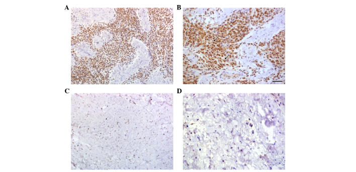
Figure 1.
Immunohistochemical analyses of SOFAT expression in gingival tissue samples. Gingival tissue samples from healthy patients stained with rabbit polyclonal IgG anti-SOFAT at (A) magnification, ×200 and (B) magnification ×400. The arrows indicate plasma cells. (C) Gingival tissue samples from patients with chronic periodontitis stained with rabbit polyclonal IgG anti-SOFAT, observed at (C) magnification, ×200 and (D) magnification, ×400. SOFAT, secreted osteoclastogenic factor of activated T cells.

