Figure 2.
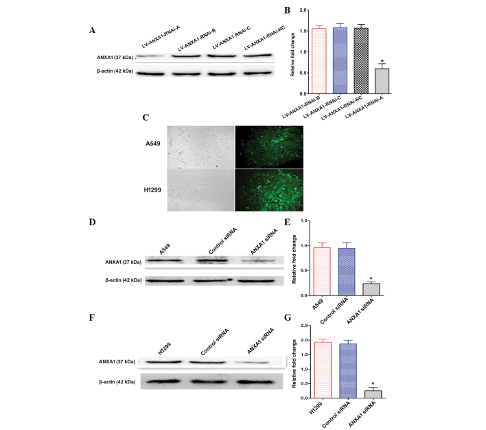
Establishment and functional analyses of annexin A1 (ANXA1) knockdown cells. (A and B) Total cellular proteins were extracted following transduction with three small interfering (si)RNA vectors. The most efficient recombinant vector was selected for further experimentation: LV-ANXA1-RNAi-A. (C) After stable transduction the cells were observed under a fluorescence microscope (magnification, 100x). (D) ANXA1 protein expression was markedly inhibited in the A549 ANXA1 siRNA group, as determined by western blotting. (E) Quantification of ANXA1 protein expression in A549 cells. (F) Western blotting indicated that ANXA1 expression was markedly decreased in the H1299 ANXA1 siRNA group. (G) Quantification of ANXA1 protein expression in H1299 cells. β-actin was used as an endogenous control. Data are presented as the mean ± standard deviation for triplicate determinations. *P<0.05, compared with the control siRNA and untreated groups.

