
Fig. 8.
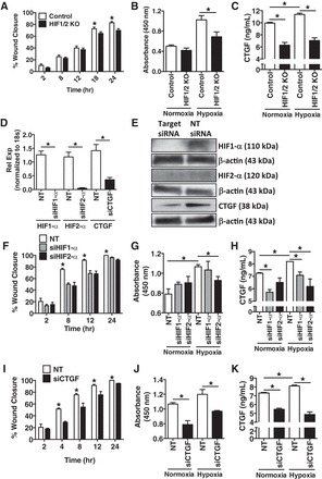
Exposure of pulmonary artery smooth muscle cells (PASMCs) to hypoxic conditioned media (CM) from primary isolated pulmonary microvascular endothelial cells (PMVECs) of control and HIF1/2 KO mice demonstrates a decrease in PASMC wound healing and cellular proliferation, to a similar degree as individual knockdown of HIF1-α, HIF2-α, and connective tissue growth factor (CTGF). A and B: murine PASMC exposure to hypoxia CM (O2 1%, 24 h) from primary isolated murine PMVECs demonstrates decreased wound healing by scratch assay (A) and decreased proliferation by BrdU assay (B) in HIF1/2 KO vs. control conditions. C: CM from HIF1/2 KO PMVECs exposed to hypoxia had a lower concentration of CTGF compared with control cells. D and E: Individually, HIF1-α, HIF2-α, and CTGF were knocked down in wild-type murine PMVECs by siRNA, with a decrease in expression by both real-time PCR (D) and immunoblot (E), compared with nontarget (denoted as NT, in figure) siRNA control. F–H: similar to above, PASMC exposure to CM from PMVECs with siRNA knockdown of either HIF1-α or HIF2-α, vs. nontarget (NT) siRNA, displayed a decrease in PASMC wound closure (both HIF1α and HIF2-α) (F) and PASMC proliferation rate (HIF2-α) (G). H: CTGF concentration in the PMVEC CM was lower with both HIF1-α and HIF2-α siRNA targeting. I–K: PASMC exposure to CM from PMVECs with siRNA knockdown of CTGF vs. nontarget (NT) siRNA, displayed a decrease in PASMC wound closure (I) and PASMC proliferation rate (J). K: CTGF concentration in the PMVEC CM was lower with CTGF siRNA targeting, but was not completely eliminated. n = 4 per group. *P <0.05.