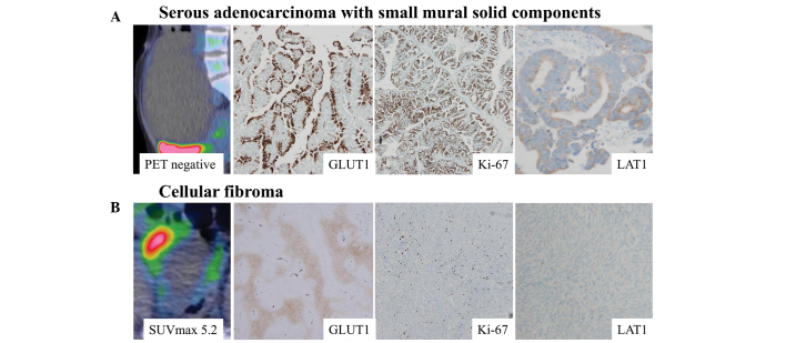
Figure 4.
Imaging and immunohistochemical findings of adenocarcinoma and fibroma. (A) A serous adenocarcinoma diagnosed as a benign lesion from preoperative PET and MRI findings. Clinicopathologically, GLUT1, Ki-67 and LAT1 expression levels were elevated. (B) An F-18 FDG-positive cellular fibroma exhibited negative Ki-67 and LAT1 expression, despite positive GLUT1 expression. Magnification, ×40. PET, positron emission tomography; MRI, magnetic resonance imaging; GLUT1, glucose transporter 1; LAT1, L-type amino acid transporter 1; FDG, fluorodeoxyglucose.

