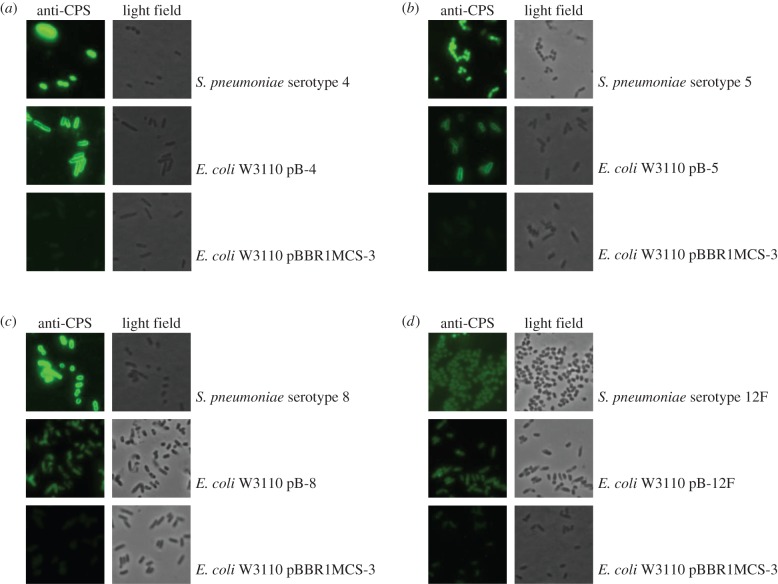
Figure 3.
Immunofluorescence microscopy of recombinantly expressed S. pneumoniae capsule loci. E. coli cells carrying the capsule expression vectors (a) pB-4, (b) pB-5, (c) pB-8 and (d) pB-12F were probed with group or type-specific anti-capsular antibody and Alexa Fluor 488 conjugated secondary antibody. Empty vector E. coli and S. pneumoniae of relevant serotype were used as controls. Images are shown at 100× magnification.

