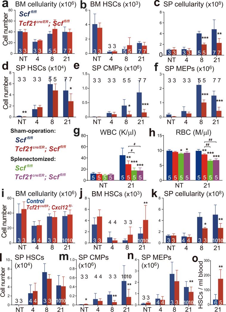
Figure 3. Tcf21-expressing stromal cells are an important source of Scf and Cxcl12 for EMH in the spleen.
a-f, Tcf21cre/ER; Scffl/fl and Scffl/fl control mice were treated with tamoxifen then examined a month later either under normal conditions (not treated, NT) or after treatment with cyclophosphamide plus 4, 8, or 21 days of G-CSF to induce EMH. The number of bone marrow cells (a) and bone marrow CD150+CD48−LSK HSCs (b) in one femur plus one tibia as well as spleen cellularity (c), and the numbers of HSCs (d), common myeloid progenitors (CMPs, e) and megakaryocyte/erythroid progenitors (MEPs, f), in the spleen. g, h, Sham-operated and splenectomized mice were treated with Cy+21d G-CSF one month after surgery: white blood cell (WBC; g) and red blood cell (RBC; h) counts. i-o, Tcf21cre/ER; Cxcl12fl/− and Cxcl12+/− or Cxcl12fl/− control mice were treated with tamoxifen then examined a month later either under normal conditions (NT) or after treatment with cyclophosphamide plus 4, 8, or 21 days of G-CSF to induce EMH. The number of bone marrow cells (i) and bone marrow HSCs (j) in one femur plus one tibia as well as spleen cellularity (k), numbers of HSCs (l), CMPs (m) and MEPs (n) in the spleen. o, Number of HSCs per ml of blood in tamoxifen-treated control and Tcf21cre/ER; Cxcl12fl/− mice after Cy+21d G-CSF. The numbers of mice per treatment are shown in each bar in each panel. All panels reflect mean±s.d. from three independent experiments. * indicates statistical significance relative to sham-operated Scffl/fl mice while # indicates statistical significance among other treatments (* or # P<0.05, ** or ## P<0.01, *** P<0.001).

