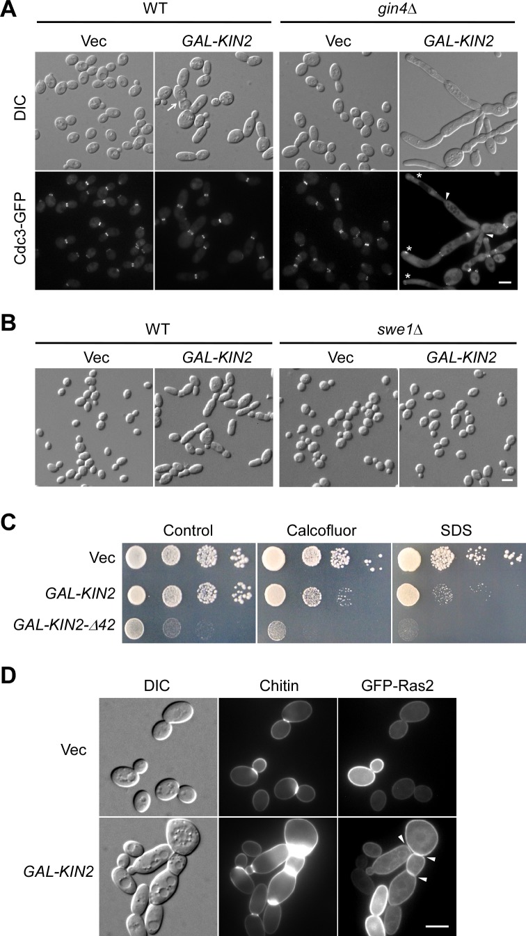
Fig 4. Overexpression of Kin2 affects septin organization and cell wall organization.
(A) Cell morphology and Cdc3-GFP localization of cells overexpressing Kin2. Cells of strain YEF473A (WT) and YEF1238 (gin4Δ) with integrated CDC3-GFP:LEU2 carrying pEGKT316 (Vec) or pEGKT316-KIN2 were grown on SRG-Ura plate at 30°C for 20 hr before imaging. (B) Morphology of wild-type and swe1Δ cells overexpressing Kin2. Cells of strain YEF473A (WT) and JGY2030 (swe1Δ) carrying pEGKT316 (Vec) or pEGKT316-KIN2 were grown on SRG-Ura plate. (C) Cells overexpressing Kin2 or Kin2-Δ42 are sensitive to calcofluor white and SDS. YEF473A cells carrying pEGKT316 (Vec), pEGKT316-KIN2, pEGKT316-KIN2-Δ42 plasmids were grown on SRG-Ura medium (control), SRG-Ura medium with 5 μg/ml calcofluor white, and SRG-Ura medium with 40 μg/ml SDS at 30°C. Pictures were taken after 6 days. (D) Chitin distribution and GFP-Ras2 localization. Cells of strain YEF473A carrying plasmids pRS315-GFP-RAS2/pEGKT316 (Vec) or pRS315-GFP-RAS2/pEGKT316-KIN2 were grown on SRG-Leu-Ura plate at 30°C for 20 hr. Chitin was stained before imaging. Bars, 5 μm.

