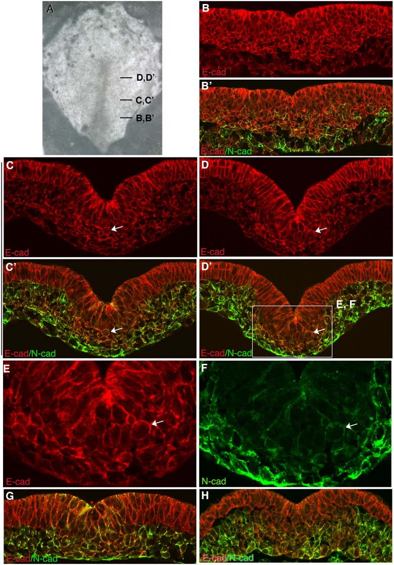
Fig 4. Localization of E-cad and/or P-cad and N-cad in transverse sections of a HH stage 3–4 chicken embryos.
(A) Brightfield image of a HH stage 3 embryo following processing for immunofluorescence localization of E-cad and/or P-cad and N-cad, showing the location of images in (B-D’). Expression of E-cad and/or P-cad (B,C,D), or E-cad and/or P-cad plus N-cad (B’, C’, D’) in transverse sections. (E, F) Higher magnification images of boxed area in (D’), showing the localization of E-cad and/or P-cad (E) and N-cad (F) as cells transition from epiblast to mesoderm. E-cad and/or P-cad are retained on the surface of cells during EMT and after cells emerge into the mesoderm. Arrows point to rounded cells in the ventral streak showing robust detection of E-cad and/or P-cad at the cell periphery. (G, H) Co localization of E-cad and/or P-cad and N-cad in transverse sections of HH stage 4 embryos, showing their heterogeneous expression between mesoderm cells.

