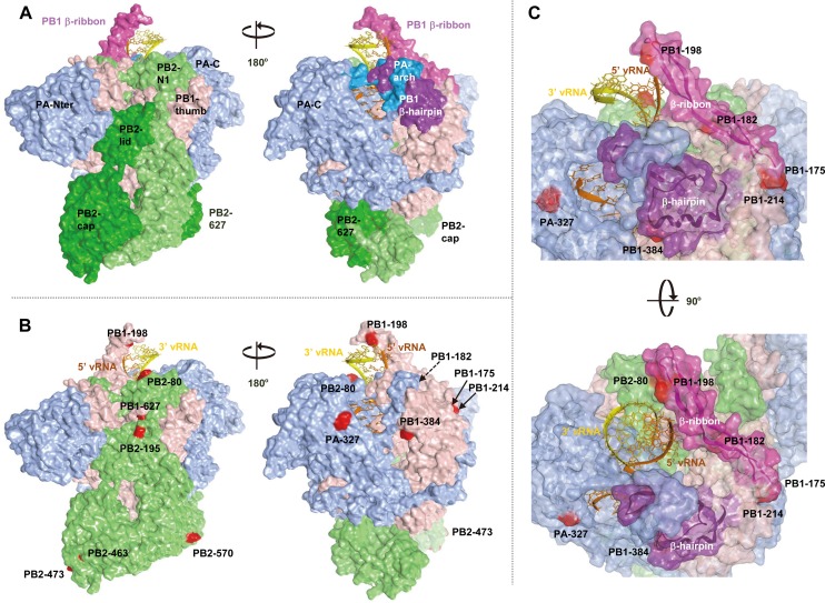
Fig 7. Structural model of the EG/D1 polymerase complex.
Structural model of the EG/D1 heterotrimeric polymerase complex bound to the vRNA promoter. (A) Surface view of the EG/D1 structure color-coded (as described below) according to the domain structure in which the mutations identified in this study were located. Left and right structures differ by 180° in orientation. (B) Surface view of the EG/D1 structure color-coded showing PB2 (light green), PB1 (pink), PA (light blue) and the mutations in this study (red). Left and right structures differ by 180° in orientation. (C) Transparent EG/D1 surface diagram showing the mutations identified in this study (red) located mainly on the PB1 β-ribbon (magenta) and β-hairpin structures (violet) sandwiching the vRNA promoter. Upper and lower structures differ by 90° in orientation.

