
Fig 2.
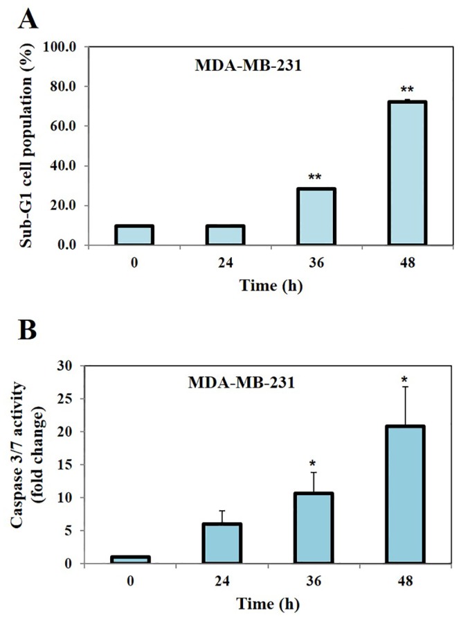
A, The analysis of cell cycle distribution following treatment with CIP was performed using flow cytometry. MDA-MB-231 cells were exposed to compound CIP (50 μM) for indicated time intervals (0, 24, 36 and 48 h), after which the cells were harvested and stained with PI. The cell distribution across the various phases of the cell cycle was analyzed with a flow cytometer. We observed that CIP induced significant apoptosis in a time dependent manner as evidenced by increased accumulation of cells in Sub-G1 phase of the cell cycle. B, MDA-MB-231 cells were exposed to compound CIP (50 μM) at indicated time intervals (0, 24, 36 and 48 h), after which they were harvested and caspase3/7 activity was measured using Caspase-Glo® 3/7 assay kit. We found that treatment of MDA-MB-231 cells with CIP caused the significant increase in the caspases-3/7 activity. * for p<0.05, ** for p<0.005.