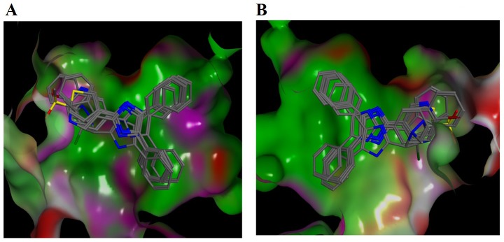
Fig 6. In silico molecular docking studies of trisubstituted imidazoles against kinase domain of Akt2:
Common binding poses of trisubstituted imidazoles towards the Akt2 kinase domain. The molecular surface of the protein is represented based on the surface polarity; green, pink and red colours show hydrophobic, polar and solvent exposed regions, respectively. For the sake of better visualisation of the binding pocket surface of Akt2, molecular surface was rendered in two panels (A and B) in which ligands were rotated 180 degrees.

