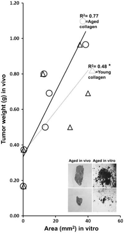
Figure 2.

Tumor weights in aged mice in vivo correlate with tumor areas in aged 3D collagen in vitro. Collagen was extracted from tail tendons of aged or young C57Bl/6 male mice (n=6 in each age group), pooled, and polymerized into 3D collagen as previously described (Damodarasamy et al. 2010). Tumors grown in aged mice in vivo and that ranged from 165 to 900 mg in size were minced into fragments. Equivalent size and weights of tumor fragments were then placed in 35 μl of 3D collagen gels derived from aged or young mice tail tendons. Tumors were allowed to grow for 5 d, and then, tumor size was determined by area calculations from digital images. The line graph shows that tumor sizes in aged 3D collagen correlated highly with their original tumor size in aged mice in vivo (R2=0.77; circles; p=not significant). In contrast, tumor sizes in young 3D collagen did not correlate with their original tumor size in aged mice in vivo (R2=0.48; triangles; *p<0.05). Representative insets show that a tumor that grew large in aged mice in vivo had robust growth in aged 3D collagen in vitro (upper panels), and a tumor that was small in aged mice in vivo grew poorly in aged 3D collagen in vitro (lower panels).
