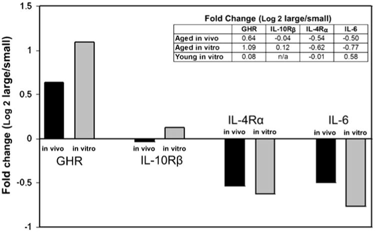
Figure 3.

Gene expression in aged mice in vivo correlates with expression in aged 3D collagen in vitro. Tumors grown in aged mice in vivo were removed and a portion immediately processed for RNA extraction using Trizol (Ambion). The other portions of the tumor were grown in aged 3D collagen or young 3D collagen in vitro, and after 5 d, cells from these tumors were isolated and processed for RNA extraction in a manner identical to that from the primary tumors. The mean fold changes (log 2) in gene expression of the three largest tumors relative to the three smallest tumors in vivo and in vitro were calculated. The bar graph shows that gene expression changes in aged mice in vivo were maintained during tumor growth in aged 3D collagen in vitro: relative expression of GHR was increased, IL-10Rβ was unchanged, and IL-4Rα and IL-6 were decreased in the larger tumors relative to the smaller tumors. The inset table shows fold change (large versus small) when comparing gene expression in tumors grown in aged mice in vivo and gene expression when the same tumors were placed in aged 3D collagen in vitro and young 3D collagen in vitro. Note that gene expression patterns were not maintained when the tumors grown in aged mice were placed in young 3D collagen (n/a=not detected).
