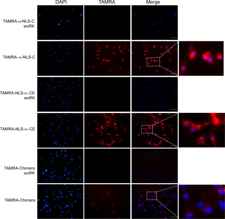
Fig 3. Designed peptides internalized and reach nuclear localization in mouse cells.
Primary cultured MEF cells were exposed for 20 min to 3 designed CPPs labeled with TAMRA at the N-terminus (TAMRA-α-NLS-C, TAMRA-NLS-α-CE and TAMRA chimera; see Table 1) or their respective control peptides (TAMRA-α-NLS-C woRK, TAMRA-NLS-α-CE woRK and TAMRA-chimera woRK; see Table 1). All peptides were used at a final concentration of 60 μM. To evaluate the co-localization of these peptides with nuclear DNA, cells were stained with DAPI after 20 min. The figure shows the fluorescence observed in DAPI, TAMRA and merged images to show the co-localization of DAPI and TAMRA signals; the squares indicate the area magnified to the right. The scale bar represents 100 μm.

