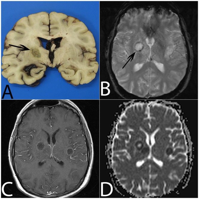
Fig 3. Aspergillosis abscess in the right thalamolenticular area due to hematogenous dissemination (patient #13).

(A) On gross examination, the lesion is non-hemorrhagic with central necrosis (arrow). (B) On T2*, the abscess is surrounded by a mild hypointense ring (arrow). (C) Gadolinium-enhanced T1W imaging shows mild annular enhancement. (D) ADC cartography shows a target-like lesion with a central high ADC value, a circular area with a low ADC value and a peripheral upper ADC value rim.
