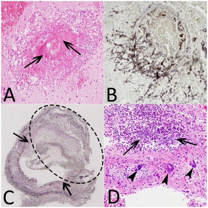
Fig 5. Histological findings (patients #9, #12, #13).

(A) Hematoxylin-eosin stain (HE) (×20), destruction of a vessel with fibrinoid necrosis (arrows). (B) Grocott methenamine silver stain (GMS) (×40), vascular wall invaded by branching septate hyphae. (C) GMS (×4), intracerebral fungal aneurysm (dotted ellipse) with the interruption of the internal elastic lamina (arrows). (D) HE (×20), aneurysm wall containing hyphae, polynuclear cells (arrows) and giant cells (arrowheads).
