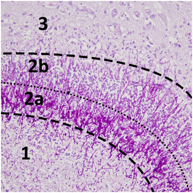
Fig 6. Histological abscess layers.

Periodic acid-Schiff stain (×20) shows distinct areas with (1) central necrosis, (2a) an intermediate dense hyphal rim, (2b) an external layer of granulation tissue and (3) edematous brain tissue. On MRI, annular enhancement after gadolinium and mild hypointense signal on T2*-weighted images correspond to layer 2a and 2b (see Fig 2C and 2D).
