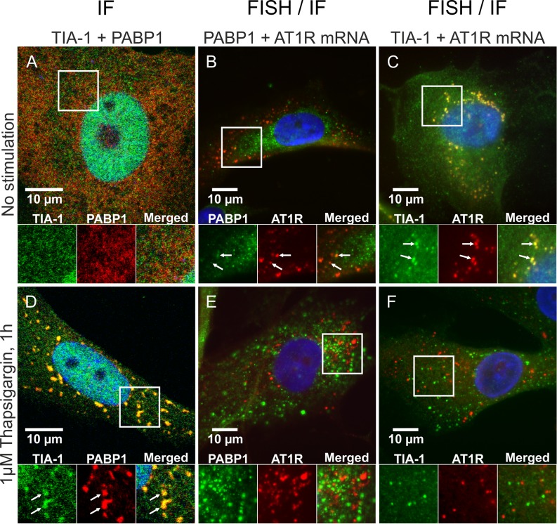
Figure 4.
AT1R mRNA dissociates from TIA-1 in response to ER stress. VSMCs were exposed to vehicle (A–C) or 1 μM thapsigargin for 1 h (D–F). After stimulations the cells were fixed and immunofluorescently stained against TIA-1 and PABP1 (A and D). Alternatively the cells were in situ hybridized with a fluorescently labeled RNA probe against AT1R mRNA and immunofluorescently stained against PABP1 (B and E) or TIA-1 (C and F). The subcellular localization of the mRNA and proteins (FISH/IF) was visualized with a fluorescence microscope whereas colocalization of the proteins (IF) was visualized with a confocal laser scanning microscope. Colocalization of the signals are indicated (arrows). The nuclear localization of TIA-1 is visible in the IF stainings (A and D) as the cells are permeabilized with 0.25% TX-100. In the FISH combined with IF stainings against TIA-1 (C and F) the nuclear localization of TIA-1 is not shown as the cells are permeabilized with 70% ethanol leaving the nuclear proteins undetected by the antibodies. The details of the staining procedures are given under ‘Materials and Methods’ section.

