Figure 3.
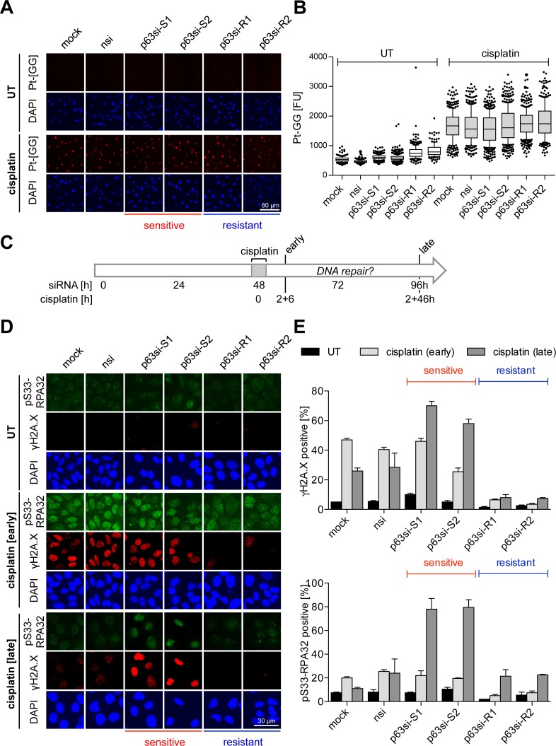
ΔNp63 regulates DNA damage repair. UT-SCC-74A were transfected with siRNAs for 48 h followed by cisplatin treatment [8 μM] for 2 h. Cells were analyzed by immunofluorescence at indicated time points. Cisplatin response is indicated as sensitive or resistant. (A and B) Detection of platinum-DNA adducts with Pt-[GG]-specific antibody (28) 6 h after cisplatin treatment. (A) Representative immunofluorescence images of Pt-[GG] adducts. DAPI: nuclear counterstain. (B) Quantification of platinum-DNA adducts from A by single-cell analysis. Box plots show fluorescence intensities [FU] of single cells, whiskers: 10th and 90th percentiles. (C–E) Time course experiment for analysis of DNA damage induction and repair. (C) Experimental setup: siRNA transfection, cisplatin treatment (for 2 h) and time points of analysis (early, 2 + 6 h and late, 2 + 46 h) are indicated. (D) Representative immunofluorescence images of DNA damage markers γH2A.X and serine 33-phosphorylated (pS33) RPA32. DAPI: nuclear counterstain. (E) Quantification of DNA damage markers from (D). Bars show mean % +SEM (n = 2) of cells positive for γH2A.X (top) and pS33-RPA32 (bottom).

