Figure 3.
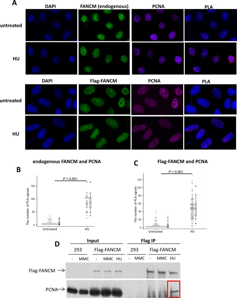
FANCM and PCNA interact in cells under replication stress. (A) Immunofluorescence images, and (B,C) quantitation of the PLA assay showing the FANCM and PCNA interaction in HeLa cells under replication stress. Cells untreated or treated with HU are indicated. FANCM was stained with either an antibody against endogenous FANCM (top 2 rows), or a Flag antibody that reacts with Flag-FANCM ectopically expressed in HeLa cells. DNA is co-stained with DAPI. In the quantitation plots each spot represents the number of PLA signals in an individual nucleus. Two independent experiments were performed for each analysis. In the experiments with exogenous FANCM a total of 144 nuclei from the untreated cells and 162 nuclei from the HU treated cells were examined. In the experiment with the Flag-FANCM a total of 157 nuclei from non-treated cells and 156 nuclei from HU treated cells were analyzed. (D) Immunoblotting shows that Flag-FANCM transfected in HEK293 cells co-immunoprecipitates with PCNA in cells treated with HU, but not in untreated cells, or in cells treated with MMC.

