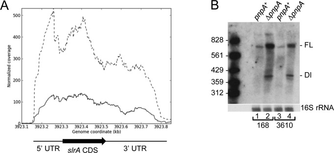
Figure 1.
Increased level of slrA mRNA in the absence of PNPase. (A) Read data from RNA-Seq analysis of the slrA gene in pnpA+ (solid line) and ΔpnpA (dashed line) strains. Genome coordinates on the X-axis; normalized reads on the Y-axis. Regions of the slrA transcription unit are indicated below the data. (B) Northern blot analysis of slrA mRNA in Bacillus subtilis 168 and 3610 backgrounds. Ten microgram of total RNA was fractionated on a 6% denaturing polyacrylamide gel. FL, full-length mRNA; DI, decay intermediate. Marker lane contained 5′-end-labeled fragments of a TaqI digest of plasmid pSE420 (51).

