Figure 2.
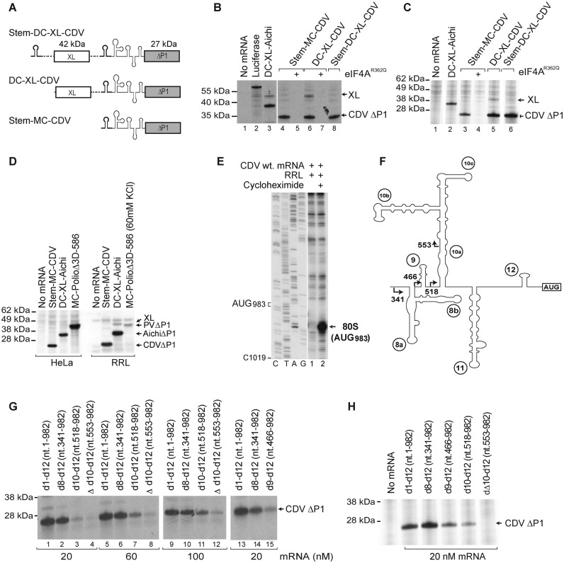
The 5’ border of the CDV 5’UTR IRES. (A) Schematic representation of monocistronic (MC) and dicistronic (DC) CDV mRNAs, with the CDV 5’UTR and a stable hairpin (thick line) placed upstream of the first and/or the second cistron. (B-D) Translational activity of the IRES in (B and D) RRL and (C and D) HeLa cell-free translation extract, and its sensitivity to inhibition by dominant-negative eIF4AR362Q. Luciferase and DC Aichivirus mRNAs (31) were used as controls for the efficiency of translation. (E) Toe-print analysis of 80S ribosomes formed on wt CDV MC mRNA in RRL in the presence of cycloheximide. (F) Model of the IRES, with arrows indicating the borders of 5’-terminal truncations of the 5’UTR. (G and H) Effect of these truncations, introduced into Stem-MC mRNA, on IRES activity in (G) RRL and (H) HeLa cell-free translation extract.

