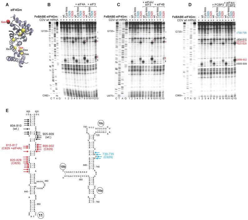
Figure 5.
Interaction of the CDV IRES with eIF4G. (A) Ribbon diagram of eIF4Gm (PDB: 1HU3) with yellow spheres indicating native (C819, C821, C847, C919, C934 and C936) and red spheres showing introduced (C829, C929) cysteines. (B–D) Primer extension analysis of directed hydroxyl radical cleavage of wt CDV mRNA from Fe(II)-tethered wt eIF4Gm, eIF4GmT829C and eIF4GmD929DC in the presence and absence of (B) eIF4A or eIF3, (C) eIF4A/eIF3 or eIF4B and (D) PCBP2 or eIF4A/PCBP2. (E) Sites of hydroxyl radical cleavage from eIF4GmT929C (blue arrows), eIF4GmD829DC (red arrows) and wt eIF4Gm (black arrows) mapped onto models of IRES d11 and the apex of d10.

