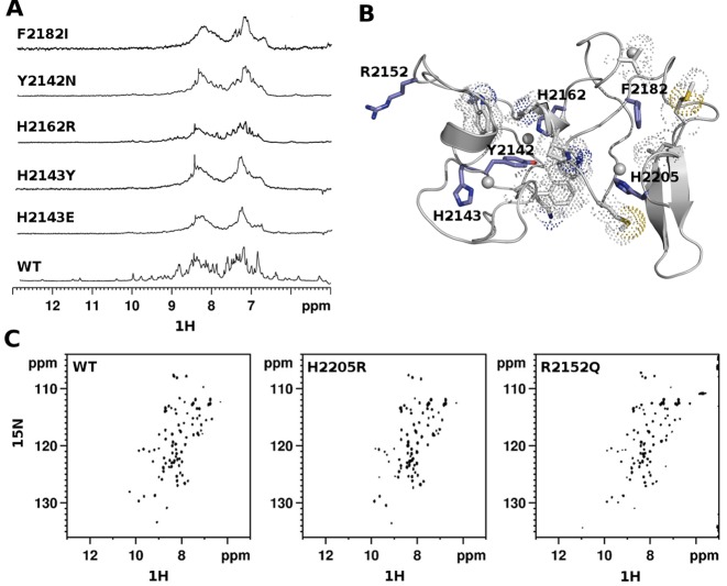
Figure 2.
Structural effects of Sotos syndrome mutations. (A) 1D 1H spectra (amide region) of wild-type and pathological mutants of PHDvC5HCHNSD1. (B) Cartoon representation of PHDVC5HCHNSD1 (gray). The side chains of pathological point mutations are shown in blue sticks, residues involved in hydrophobic contacts are shown in grey sticks with dotted space-filled representation. (C) 2D 1H–15N HSQC spectra (amide region) of wild-type and pathological mutants of PHDvC5HCHNSD1.

