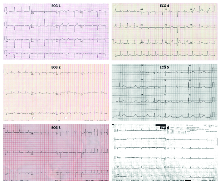
Figure 1.
ECG findings and patient presentations.
ECG 1: Left axis deviation, single atrial ectopic beat. ’40-year-old female with flutters.’
ECG 2: ST elevation. ’55-year-old male, syncope at the gym’ (RED FLAG).
ECG 3: Atrial fibrillation. ’75-year-old female, diabetic, no symptoms.’
ECG 4: Normal ECG. ‘50-year-old female, sudden onset and offset regular palpitations.’
ECG 5: Prolonged QT interval. ’16-year-old female, blackout in school assembly’ (RED FLAG).
ECG 6: Poor-quality ECG, sinus rhythm, erroneous automated report of AF. ’81-year-old female, breathless.’

