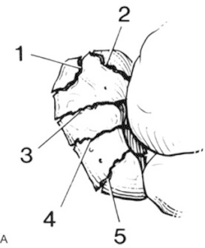
Fig. 2.

Cooney (Mayo) divided scaphoid fractures into fractures of the distal tubercle (1), distal intra-articular surface (2), distal third (3), waist (4), and proximal pole (5). Fracture location influenced both tendency and time frame for healing. (Reprinted, with permission covered by STM guidelines, from Cooney et al.13)
