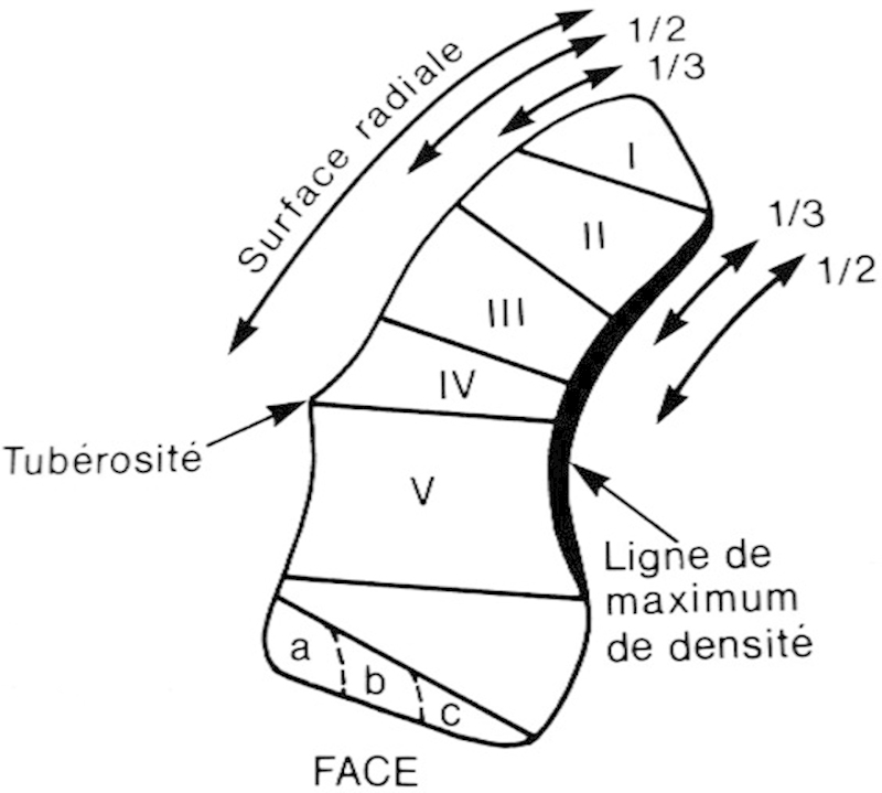
Fig. 3.

Schernberg distinguished six fracture types (I–VI) ranging from the proximal pole to the distal tubercle using the lateral tuberosity and the radial and medial articular surfaces as references. Distal tubercle fractures were further divided into small (a), intermediate (b), or large (c) fragments, and were considered likely to heal successfully, contrary to proximal fractures. (Reprinted, with permission of authors, from Schernberg et al.21)
