Figure 1.

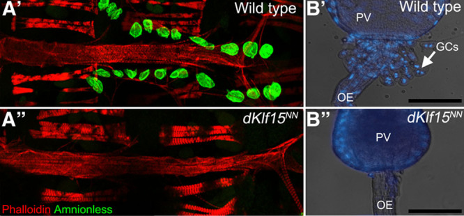
Loss of pericardial nephrocytes and garland cells in dKlf15NN mutants. A, Adult control (w1118; A′) and dKlf15NN mutant flies (A″) were dissected and the heart fixed and stained with phalloidin to visualize the heart’s actin cytoskeleton and antibodies to the pericardial nephrocyte marker Amnionless (CG11592). All pericardial nephrocytes fail to differentiate in the mutants, undergoing attrition during late larval development so that by adulthood there are none. B, Third instar larvae were dissected and the garland cells visualized after staining with Hoechst. In control flies (w1118; B′) garland cells (GCs) are binucleate and situated at the interface between the proventriculus (PV) and esophagus (OE). In contrast, the garland cells fail to develop normally and are lost in the dKlf15NN mutants (B″). Scale bar, 100 μm.
