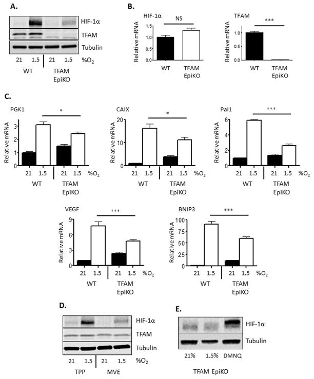
Figure 1.
Mitochondrial generation of ROS is required for hypoxic induction of HIF-1α protein and target gene expression in mouse epidermal keratinocytes. (A) Representative Western blot analysis of HIF-1α and TFAM protein levels in primary mouse keratinocytes isolated from wild-type and TFAM EpiKO mice. Cells were exposed to normoxia (21% O2) or hypoxia (1.5% O2) for 4 hours. (B) Real Time PCR analysis of HIF-1α and TFAM mRNA expression in primary mouse keratinocytes isolated from wild-type and TFAM EpiKO mice. (C) Real Time PCR analysis of PGK1, CAIX, Pai1, VEGF, and BNIP3 mRNA expression in primary mouse keratinocytes isolated from wild-type and TFAM EpiKO mice. Cells were exposed to normoxia or hypoxia for 4 hours. (D) Representative Western blot analysis of HIF-1α protein levels in primary wild-type mouse keratinocytes after exposure to normoxia or hypoxia for 4 hours. Cells were treated with mitochondria-targeted vitamin E (MVE) or the control compound methyl-triphenylphosphonium (TPP; mitochondria-targeting moiety lacking antioxidant activity). (E) Representative Western blot analysis of HIF-1α protein levels in TFAM EpiKO primary mouse keratinocytes after exposure to normoxia, hypoxia, or 2,3-dimethoxy-4-naphthoquinone (DMNQ) for 4 hours. Charts represent means +/− SEM. N=4 independent keratinocyte preparations per genotype. mRNA expression was normalized to RPL19 levels. Significance was determined by (B) Students T-test or (C) one way ANOVA using Bonferonni’s post-test.

