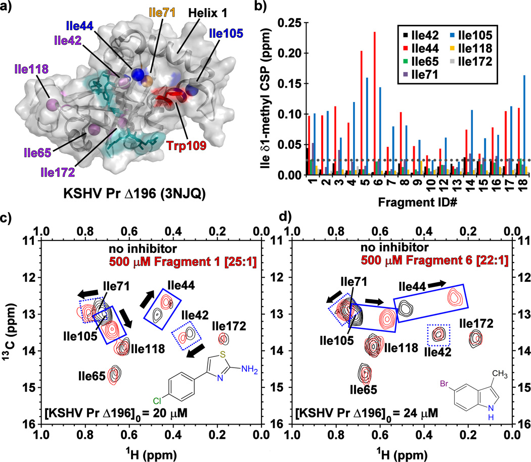
Figure 2.
(a) The structure of monomeric KSHV Pr Δ196 (PDB: 3NJQ), with the catalytic (cyan), hot spot Trp109 (red), and surface exposed Ile44 and Ile105 (blue) residues. Helix 1, Ile71 (orange) and the remaining isoleucine residues (purple) also indicated. (b) The chemical shift perturbations (CSPs) of the isoleucine δ1-methyl groups in the presence of the Table 1 fragments. The gray dotted line represents a CSP threshold of 0.025 ppm. Representative 13C/1H-HSQC spectral overlays of selectively [13C-methyl] isoleucine labeled KSHV Pr Δ196 in the absence (black) and presence (red) of Fragments (c) 1 and (d) 6.

