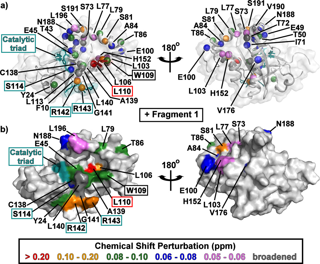
Figure 4.
The structure of monomeric KSHV Pr Δ196 (PDB: 3NJQ) with the 15N/1HN-HSQC chemical shift perturbations for Fragment 1 indicated by color. Backbone amide resonances which displayed peak broadening upon addition of fragments are indicated in dark gray. Amide backbone nitrogen atoms are shown as colored spheres in (a), while surfaces are displayed in (b). The catalytic triad (His46, Ser114, and His134) and oxyanion hole (Arg142 and Arg143) residues are highlighted in cyan. Left and right structures are rotated 180° about the vertical axis. CSP mapping results for Fragment 6 are displayed in Figure S4.

