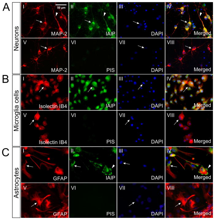
Fig. 2.
IAIPs immunoreactivity in cultured rat cortical neurons, microglial cells and astrocytes. (A): Double immune-staining for MAP-2 (AI) and IAIPs (AII) shows that IAIPs were confined to neurons [cell body (arrow)] and dendrites (arrowhead), AIV). (B): Double immune-staining for Isolectin IB4 (BI, arrow) and IAIPs (BII, arrow) demonstrated the presence of IAIPs in the cytoplasm of microglial cells (BIV, arrow). (C): Double staining for GFAP (CI, arrow) and IAIPs (CII, arrow) revealed a perinuclear localization of IAIPs in astrocytes (CIV, arrow). Localization of IAIPs in the end-feet area of astrocytes was observed (CII and CIV, arrowhead). PIS negative staining and DAPI counter-staining were performed for each experiments. Bars: 50 μm.

