Fig. 3.
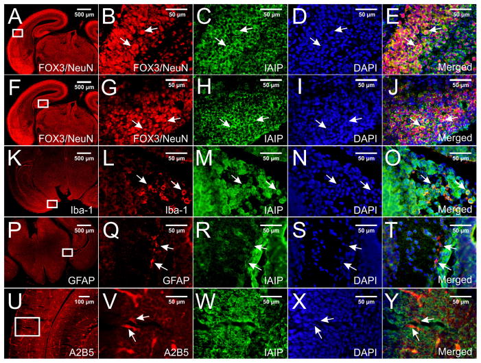
Double immune-staining with pR-21 and with specific brain cell markers in paraffin-embedded E18 mouse brain sections. IAIPs immunoreactive signals were detected in different brain regions including cortex and hippocampus. Figs. 3E and 3J show co-localization of IAIPs with FOX3/NeuN (Figs. 3B and G) in neurons in the entorhinal cortex and hippocampus, respectively. Figs. 3O and 3T show co-localization of IAIPs with Iba-1 (L) and GFAP (Q) in the microglial cells and astrocytes in the area of medial hippocampus, respectively. A2B5-positive (U and V) glial progenitors were not found to co-localize with IAIP (W and Y). Specific IAIPs staining was mainly localized to cytoplasm and perinuclear of cells (E, J, O, T arrows). DAPI was used as a counterstaining (D, I, N, S, X).

