Figure 7.
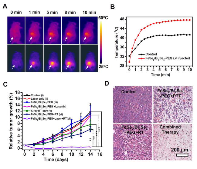
In vivo combined photothermal & radiation therapy. (A) IR thermal images of 4T1 tumor-bear mice without (upper row) or with i.v. injection of FeSe2/Bi2Se3-PEG (lower row, dose = 20 mg/kg, irradiated at 24 h p.i.), under the 808-nm laser irradiation taken at different time intervals. The laser power density was 0.5 W/cm2. (B) Temperature changes of tumors monitored by the IR thermal camera during laser irradiation. (C) The tumor volume growth curves of mice after various treatments (5 mice for each group). Group i: Untreated control; Group ii: NIR laser only; Group iii: FeSe2/Bi2Se3-PEG; Group iv: FeSe2/Bi2Se3-PEG + NIR; Group v: X-ray RT alone; Group vi: FeSe2/Bi2Se3-PEG + RT; Group vii: FeSe2/Bi2Se3-PEG +NIR + RT. PTT was conducted by the 808-nm at 0.5 W/cm2 for 10 min, while the irradiation dose of RT was 4 Gy. Error bars were based on standard error of the mean (SEM). (D) Micrographs of H&E stained tumor slices from different groups of mice treated with PBS, FeSe2/Bi2Se3-PEG+NIR, FeSe2/Bi2Se3-PEG+RT, and FeSe2/Bi2Se3-PEG+RT+NIR. The tumors were harvested 2 days after treatments were conducted.

