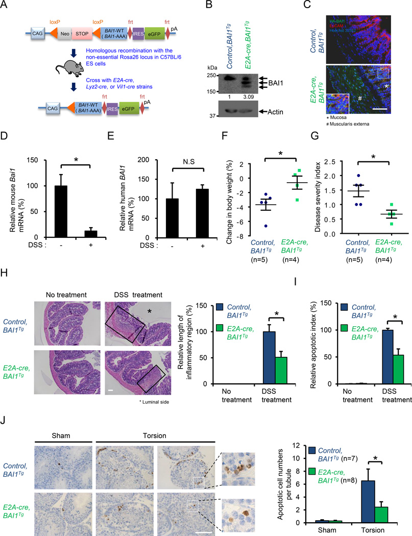
Figure 3. Transgenic overexpression of BAI1 attenuates acute colonic inflammation.
(A) Schematic for generation of BAI1Tg mice expressing human BAI1, via cre-mediated deletion of the STOP cassette preceding the transgenes. E2A-cre yielded global BAI1Tg mice, while Lyz2-cre (myeloid) and Vil1-cre (intestinal epithelial cells) directed tissue specific Bai1Tg expression.
(B) Detection of HA-BAI1 in the colon of control and E2A-cre, BAI1Tg mice via immunoblotting. All three bands represent BAI1 (due to differential glycosylation).
(C) Colon from control and E2A-cre, BAI1Tg mice were analyzed for HA-tag on transgenic BAI1 (green), the epithelial cell marker EpCAM (red) and Hoechst 33342 (blue). Scale bars=100 µm.
(D and E) Mouse Bai1 mRNA and transgenic human BAI1 mRNA in purified gut epithelial cells from E2A-cre, BAI1Tg mice after DSS (3%) treatment (No treatment: n=3, 3M; DSS: n=3, 3M).
(F and G) Change in body weight and disease severity index after DSS (3%) treatment in control (n=5; 5M) and E2A-cre, BAI1Tg (n=4; 4M) mice.
(H, I) H&E staining and apoptotic cell numbers in proximal colon from control and E2A-cre, BAI1Tg mice before and after DSS (3%) treatment (No treatment: n=5 for control, n=3 for E2A-cre, BAI1Tg; DSS treatment: n=6; 6F for control, n=4; 4F for E2A-cre, BAI1Tg). Scale bars=100µm.
(J) Cleaved caspase-3 (CC3) staining of testes from control and E2A-cre, BAI1Tg mice after sham treatment or testicular torsion (n=7; 7M for control, n=8; 8M for E2A-cre, BAI1Tg). Scale bars=50µm.
Data are representative of at least two or three independent experiments. Error bars indicate s.e.m. *P<0.05. M, male. F, female. See also Figure S3.

