. 2016 Mar 31;43(1):15–25. doi: 10.5653/cerm.2016.43.1.15

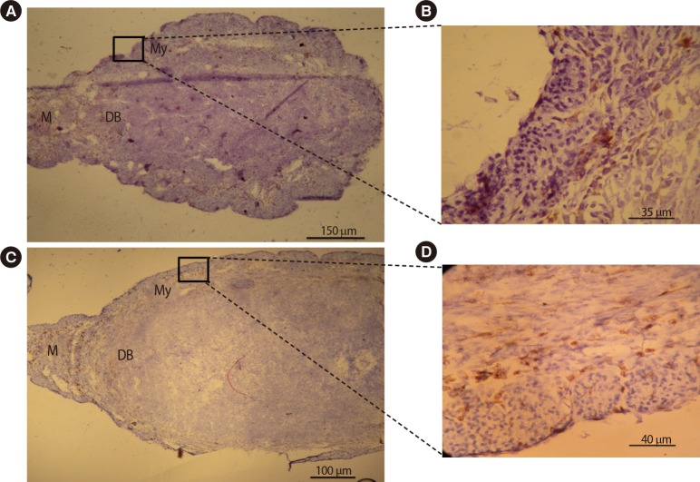
Figure 4. Immunohistochemical comparative analysis of macrophages in the uteri of pregnant BALB/c mice on gestational day 7.5. Cryosections were prepared from the uteri of pregnant BALB/c mice and stained with F4/80 antibody. Tissues were also counterstained with hematoxylin. Positive cells were counted and expressed as a percentage of the total number of nucleated cells. Macrophages were present in the myometrium, mesometrium, and decidua basalis. The frequency of uterine macrophages significantly decreased in pregnant BALB/c mice treated with DEX (A, B) when compared with pregnant BALB/c mice treated with PBS (C, D). DEX, dexamethasone; PBS, phosphate-buffered saline; M, mesometrial side; DB, decidual basalis; My, myometrium.

Figure 4