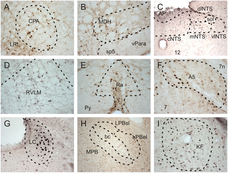
Figure 2.
Brightfield photomicrographs showing Fos-positive neurons in selected brainstem nuclei of repetitively diving rats in which the AEN had been bilaterally sectioned. Fos-positive neurons were present in the (A) caudal pressor area (CPA), (D) rostral ventrolateral medulla (RVLM), (E) Raphe nuclei (Ra), (F) A5 noradrenergic region, (G) Locus Coeruleus (LC), and (I) Kölliker Fuse region (KF). (B) Fos-positive neurons were present in the ventral tip of the superficial laminae of the medullary dorsal horn (MDH), as well as in the ventral paratrigeminal nuclei located within the spinal trigeminal tract (sp5). (C) Within the nucleus tractus solitarius at a level just caudal to the obex, Fos-positive neurons were present in the commissural (cNTS), medial (mNTS), and dorsolateral subnuclei (dlNTS), and to a lesser extent within the ventrolateral subnuclei (vlNTS). Within the parabrachial region (H), some Fos-positive neurons were found within the medial parabrachial (MPB) and external lateral subregion of the parabrachial subnuclus (LPB el), while very few Fos-positive neurons were found within the superior lateral subregion of the lateral parabrachial subnuclei (LPBsl). (B,H) at 10X; all other panels at 20X.

