
Figure 1.
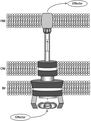
Organization of the Type III secretion system. Cartoon of the T3SS from Shigella is shown with the basal body spanning the inner membrane (IM) and outer membrane (OM) of the pathogen and the external needle inserting into the host membrane (HM). The Spa47 hexamer is depicted at the base of the needle assembly. Arrows show the path of the effector protein passage from the pathogen to the host through the apparatus.