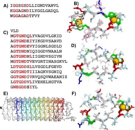
Figure 5.

Modeling the M. primoryensis ice‐binding protein (region IV) (MpAFP_RIV). A) Sequence and B) crystal structure of the repetitive β‐roll domain of P. aeruginosa alkaline protease. C) Sequence and D) modeled structure of the repetitive domain of the MpAFP_RIV. E) Peptide backbone (sticks) and calcium ion (spheres) alignment of MpAFP_RIV crystal structure (gray) and model (rainbow). F) MpAFP_RIV crystal structure. For A and C red letters indicate most repetitive residues. For B, D, and F, acidic residues are in red, basic residues are in blue, polar residues are in light cyan, aromatic residues are in light pink, and Gly are green, all other non‐polar residues are in gray. Calcium ions are gold spheres.
