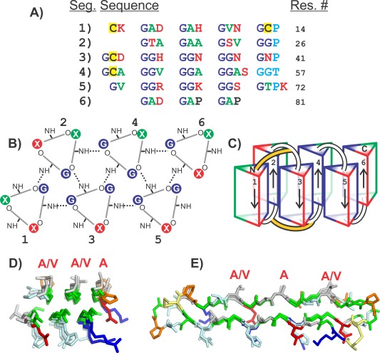
Figure 8.

Modeling the snow flea antifreeze protein (sfAFP). A) SfAFP sequence separated into six segments. Green letters are residues with small non‐polar side chains (Ala, Val). Red letters are charged or hydrophilic residues (Arg, Lys, Asp, Asn, Ser, Thr, His). Dark blue letters are Gly. Cyan letters are the residues that disturb the PPII helix. Yellow backgrounds are Cys. B) Top view of PPII helices showing hydrogen bonding pattern between coils. Segment numbers are labeled 1–6. Hydrogen bonds are shown as dotted lines. Residue positions are colored the same as in A. C) Side view of PPII bundles showing direction of each segment and disulfide bonding positions (yellow connections). D) Top view and E) side view of crystal structure of SfAFP. Structures in D and E are colored the same as in Figure 5. Additionally Cys are yellow and Pro are orange. Residues labeled with red letters are on the ice‐binding site.
