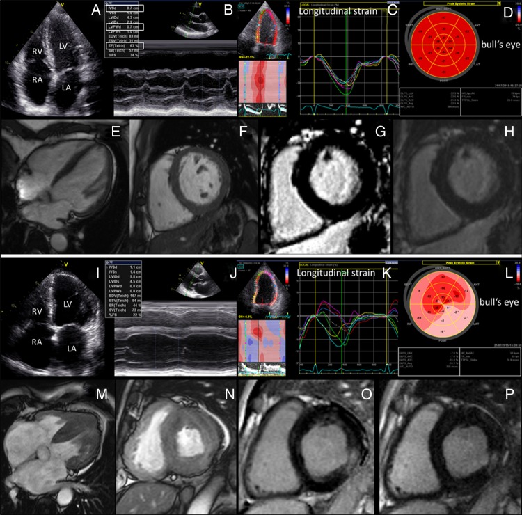
Figure 1.
Cardiac morphology and function of a representative D313Y patient versus a patient with classical Fabry disease and advanced Fabry cardiomyopathy. (A–H) In D313Y patients (participant #3), no signs of cardiac hypertrophy or diastolic dysfunction can be seen. (I–P) Fabry cardiomyopathy in respective genotypes is characterised by advanced hypertrophic thickening of the interventricular septum and the left ventricular posterior wall with left atrial dilation and reduced left ventricular ejection fraction in advanced stages. Moreover, 2D-speckle tracking reveals reduced longitudinal strain (K) with extensive replacement fibrosis visualised by bull's eye in loco typico for Fabry cardiomyopathy (L). Morphologic and LGE cardiac MRI illustrate the difference in cardiac wall thickness between D313Y patients without cardiac involvement (E–H) compared to patients with advanced Fabry cardiomyopathy (M–P).

