
Figure 1.
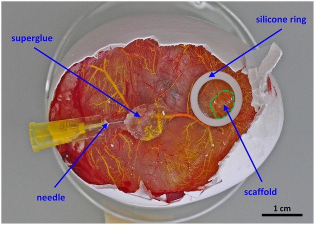
Setup for the perfusion of the chorioallantoic membrane vasculature. The scaffold is placed in the middle of a silicone ring and cultured for 7 days in ovo. For proper perfusion a 30G ½” needle is fixed with a drop of superglue before MicroFil® (yellow) injection.