
Figure 2.
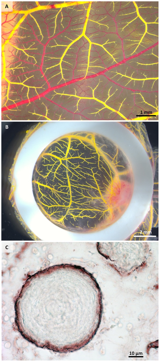
Macroscopical and microscopical view of perfused samples. (A) Perfused (yellow) and non-perfused (red) vessels after one injection showing incomplete perfusion. (B) Exemplary sample after two injections of MicroFil®. (C) Histological section of a perfused sample showing vessels (brown) containing MicroFil®.