
Figure 3.
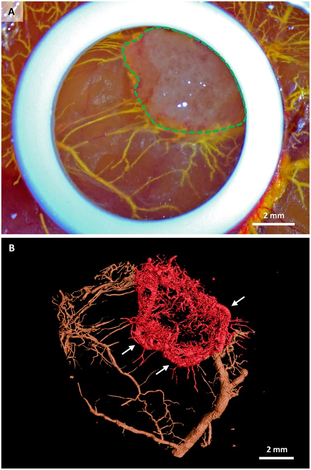
Stem cell-seeded scaffolds perfused with MicroFil® after their in ovo culture. (A) Macroscopic view of a scaffold (green dashed line) immediately after perfusion with yellow MicroFil®. The silicone ring is seen in white, vasculature in yellow. (B) Microcomputed tomography image of the vascular cast within the scaffold (red color). Vasculature outside the scaffold area is marked in brown. New capillaries are sprouting from one large vessel that encircles the scaffold (white arrows).