Figure 1.
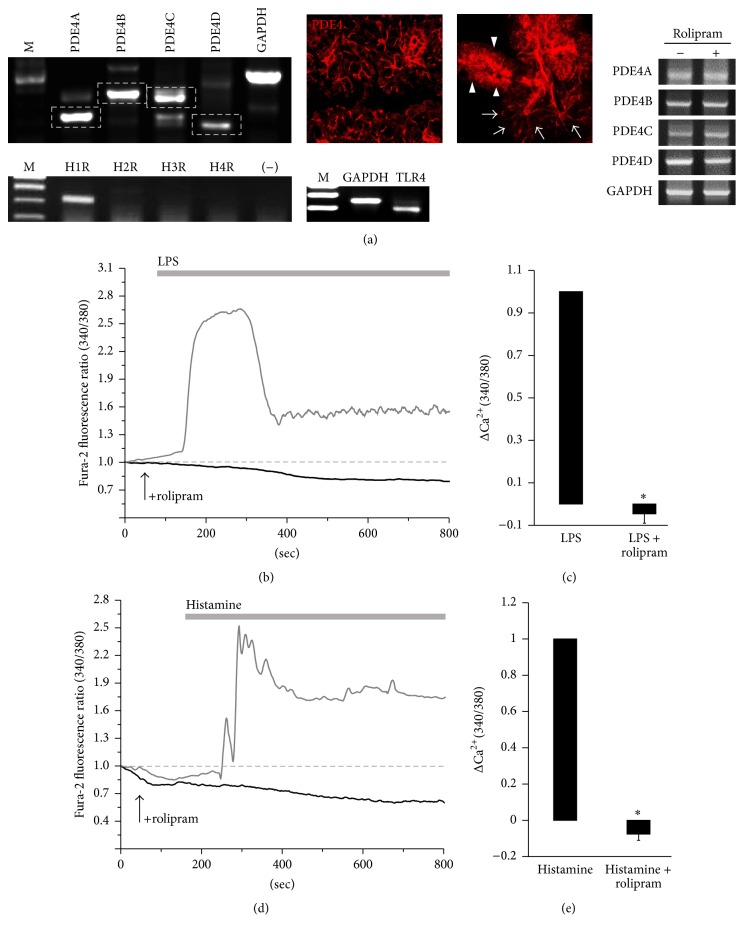
Rolipram inhibits LPS- and histamine-induced [Ca2+]i signaling in mouse SMG acinar cells. (a) mRNA expression of PDE4 subfamily 4A through 4D and localization of PDE4 (red) in SMG tissue (left) and isolated cells (right). Arrow heads (duct) and arrows (acini). mRNA expression of TLR4 and histamine receptors (H1R to H4R) in SMG cells and expression of PDE4A through 4D in the presence of rolipram. (b) Changes in [Ca2+]i induced by 20 μg/mL LPS (gray trace) and pretreatment with 10 μg/mL rolipram (black trace including arrow which indicates rolipram-stimulated time course) in SMG acinar cells. The traces are averaged trace (n = 4). The upper bars indicate the extracellular solutions applied to the cells. (c) Analysis of LPS-induced maximum [Ca2+]i peak as determined using R340/380 fluorescence ratios (∗ P < 0.01) and means ± SEMs. (d) Changes in [Ca2+]i induced by 100 μM histamine (gray trace) and pretreatment with 10 μg/mL rolipram (black trace including arrow which indicates rolipram-stimulated time course) in SMG acinar cells. The traces are averaged trace (n = 3). The upper bars indicate the extracellular solutions applied to the cells. (e) Analysis of histamine-induced maximum [Ca2+]i peak as determined using R340/380 fluorescence ratios (∗ P < 0.01) and mean ± SEMs.

