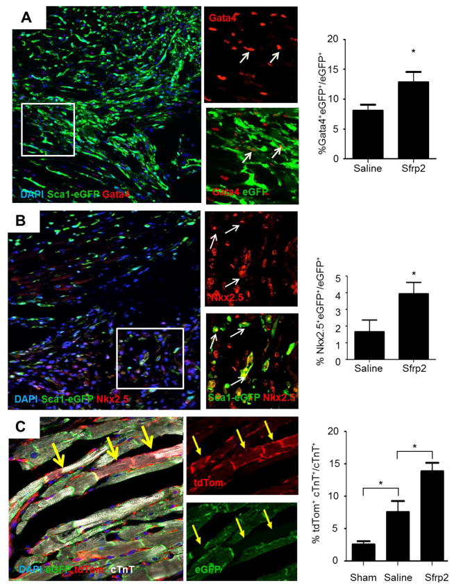
Figure 1. Sfrp2 promotes differentiation of CPCs in vivo.
(A and B) Representative images and quantification of (A) Sca1-eGFP+ and Gata4+ cells and (B) Sca1-eGFP+ and Nkx2.5+ cells 7 days after ischemia-reperfusion injury (arrows point to Sca1-eGFP+Gata4+ and Sca1-eGFP+Nkx2.5+ cells respectively); n=7 saline, n=9 Sfrp2. * P≤0.05. (C) Representative images of immunofluorescence staining of newly formed tdTomato+cTnT+ cardiomyocytes in the infarct border zone of Sfrp2 treated heart with quantification. *P<0.05, compared to PBS treated animals; n=6 Sham, n=5 PBS, n=6 Sfrp2.

