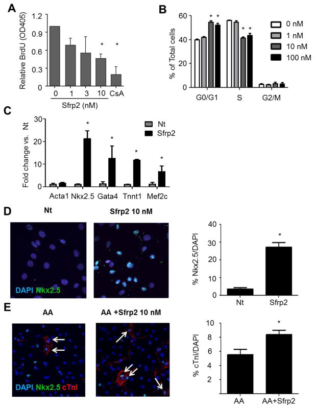
Figure 2. Sfrp2 primes CPC for cardiac differentiation.
(A) BrdU incorporation ELISA of CPCs treated for 16 hours with Sfrp2. Cyclosporin A (CsA) was used as control for decreased proliferation. Data shown as mean ± SE of 5 independent experiments. *P<0.01 compared to no treatment; n=6.
(B) FACS analysis of CPCs cultured for 8 hours in the presence or absence of Sfrp2. Ploidy was identified by 7-AAD (DNA) staining and S-phase was identified by BrdU. Data from a representative experiment are presented as mean ± SD (n=3).* P<0.05, compared to no treatment.
(C) qRT-PCR analysis for cardiac markers in non-treated (Nt) and CPCs treated with 10nM Sfrp2 for 10 days. Relative mRNA levels are expressed as fold change over control Nt cells. Data shown as mean ± SE of 4 independent experiments. * P<0.05; n=4.
(D) Representative immunofluorescence staining images for nuclear Nkx2.5 in CPCs after 14 days of continuous treatment with 10nM of Sfrp2. Data shown as mean ± SE. *P<0.05 compared to Nt; 5 images x n=3.
(E) Representative immunofluorescence staining images for cTnI in CPCs after 21 days of continuous treatment with ascorbic acid (AA) with or without 10 nM Sfrp2. Data shown as mean ± SE. *P<0.05, compared to Nt: 4–5 images x n=3. In D and E arrows denote positive cells.

