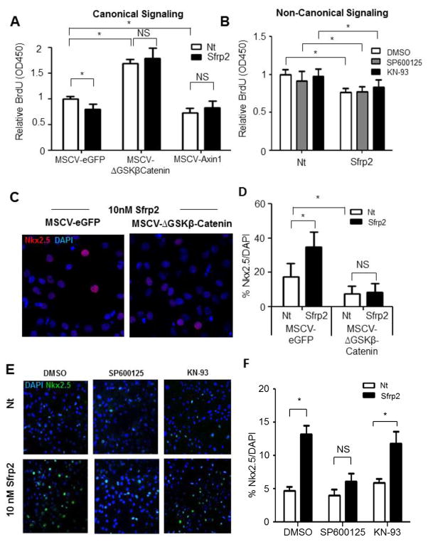
Figure 4. The effects of Sfrp2 on CPC proliferation and differentiation are mediated by inhibition of canonical and activation of non-canonical Wnt pathways.
(A) BrdU ELISA in CPCs transduced with constitutively β-Catenin (MSCV-ΔGSKβCatenin) and treated for 16 hours with 10 nM Sfrp2. MSCV-eGFP transduced cells were used as negative control. MSCV-Axin transduced cells with constitutive inhibition of β-Catenin served as positive controls. Data shown as mean ± SD. * P<0.05; n=6.
(B) BrdU ELISA of CPCs treated with 10 nM Sfrp2 for 16 hours in the presence or absence of JNK inhibitor SP600125 or CAMKII inhibitor KN-93. Data shown as mean ± SD. * P<0.05; n=6.
(C) Immunofluorescence staining for Nkx2.5 in CPCs transduced with constitutively active β-Catenin (MSCV-ΔGSKβCatenin) and treated for 14 days with 10 nM Sfrp2. MSCV-eGFP transduced cells were used as negative control. *P<0.05; 5 images x n=3.
(D) Quantitative analysis of (C).
(E) Immunofluorescence staining for Nkx2.5 after 14 days continuous treatment with Sfrp2 in the presence or absence of JNK inhibitor SP600125 or CamKII inhibitor KN-93. DMSO was used as a vehicle.
(F) Quantitative analysis of Figure 4E. Data shown as mean ± SE. *P<0.05; 5 images x n=3, analysis performed on data from 3 independent experiments. Nt: No treatment; NS: Not significant.

