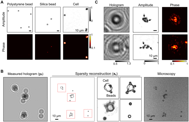
Figure 7. High resolution reconstruction of biological samples.
(A) Objects with different optical transmittance and phase contrasts were imaged: polystyrene bead, silica bead, and a mammalian cell. The sparsity algorithm recovered the optical signature, allowing for the object identification. (B) Cancer cells were labeled with polystyrene beads, and imaged by a LDIH system (left). The sparsity algorithm recovered both the amplitude and the phase information of the sample (middle). Cells and beads could be differentiated from the size as well as the optical contrast (inset). The reconstructed image was comparable to the reference image acquired by a bright field microscope with a 100x objective. (C) The sparsity algorithm was applied to detect intracellular targets. Cell nucleus was stained with a chromophore to enhance the optical contrast. The reconstructed LDIH image shows high contrast between the cell nucleus and cytosol indicated by arrows.

