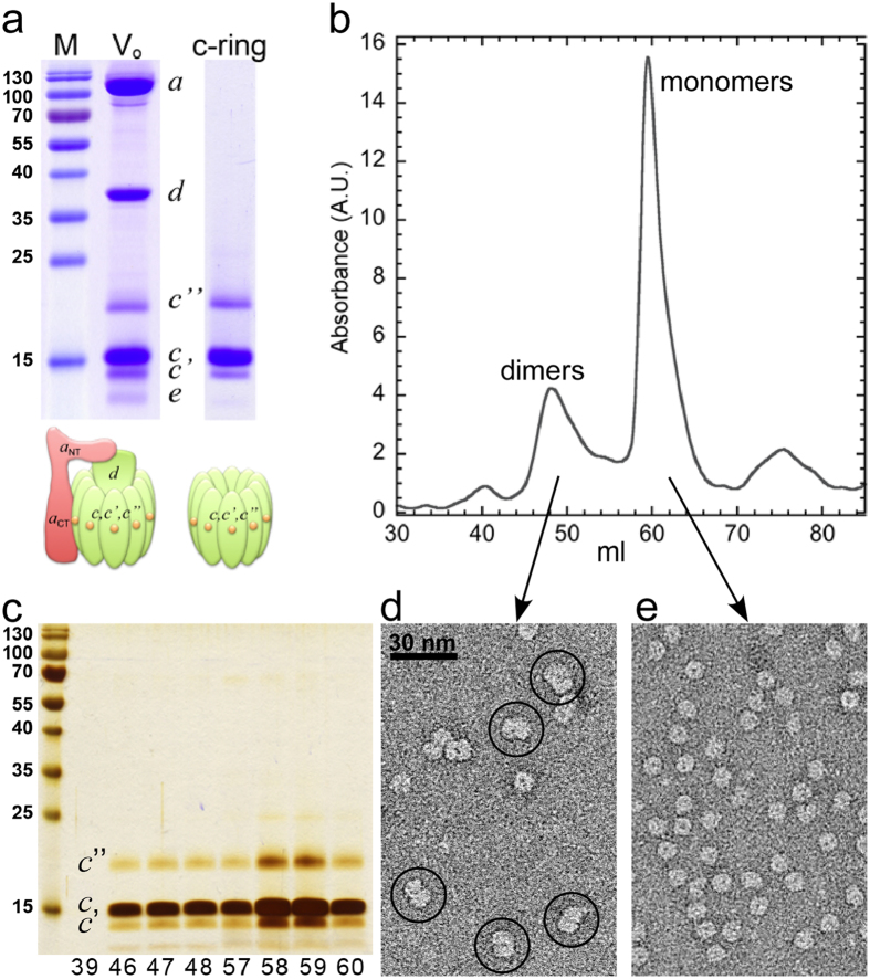
Figure 2. Purification and structural characterization of yeast V-ATPase c-ring.
(a) SDS-PAGE of purified Vo (10 μg) as well as dialyzed and concentrated c-ring (5 μg). The gels were stained with Coomassie blue. (b) Size-exclusion chromatography (Superdex 200, 16 × 500 mm2) of c-ring (1 mg) in 0.1% DDM containing buffer. (c) Peak fractions (10 μl each) were analyzed by SDS-PAGE and silver staining. (d,e) Negative stain EM of fraction 48 and 58, respectively. Dimeric c-ring complexes are highlighted by circles in (d).

